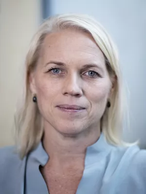
Sophia Zackrisson
Research group manager, Principal investigator, Professor, MD

Advancing breast tissue imaging with an ultrasound optical tomography (UOT) approach
Author
Editor
- Valery V. Tuchin
- Walter C.P.M. Blondel
- Zeev Zalevsky
Summary, in English
In this work an optical deep tissue imaging technique called ultrasound optical tomography (UOT) which combines laser light and ultrasound is implemented for a non-invasive lesion (tumour) characterization in breast tissue.
The experiments were performed using 794 nm laser wavelength, 6 MHz ultrasound frequency and a narrowband spectral filter material, Tm3+:LiNbO3. The measurements were carried out in 5 cm thick agar phantoms using a range of tumor mimicking inclusions of 3 different sizes.
This work is the first deep tissue imaging demonstration using UOT at tissue relevant wavelengths. Current results indicate that the UOT technique can become an important and valuable tool for lesion characterization in breast tissue.
Department/s
- LTH Profile Area: Nanoscience and Semiconductor Technology
- LTH Profile Area: Engineering Health
- LU Profile Area: Light and Materials
- LTH Profile Area: Photon Science and Technology
- NanoLund: Centre for Nanoscience
- Atomic Physics
- Chemical Physics
- Centre for Environmental and Climate Science (CEC)
- LUCC: Lund University Cancer Centre
- EpiHealth: Epidemiology for Health
- Radiology Diagnostics, Malmö
Publishing year
2024
Language
English
Pages
1301006-1301006
Publication/Series
Tissue Optics and Photonics III : PROCEEDINGS VOLUME 13010 SPIE PHOTONICS EUROPE | 7-12 APRIL 2024
Volume
13010
Document type
Conference paper
Publisher
SPIE
Topic
- Radiology and Medical Imaging
- Atom and Molecular Physics and Optics
Keywords
- breast cancer
- bio-medical imaging
- diagnosis
- ultrasound optical tomography
- deep tissue imaging
- acousto-optic effect
- spectral-hole-burning filters
- rare-earth-ion-doped crystals
Conference name
SPIE Photonics Europe, 2024, Strasbourg, France
Conference date
2024-06-18
Conference place
Strasbourg, France
Status
Published
Research group
- Radiology Diagnostics, Malmö

